Knee Muscle Anatomy Mri / Mri anatomy of knee Dr. Muhammad Bin Zulfiqar. Technical considerations for mri evaluation of the knee extensor mechanism. 12 photos of the knee muscle anatomy mri. General anatomy and musculoskeletal system. View of the anatomical labels. Tendons attach the muscles to each other.
Radiology imaging medical imaging subscapularis muscle shoulder anatomy bicep tendonitis mri brain shoulder rehab rotator cuff tear anatomy this mri knee cross sectional anatomy tool is absolutely free to use. Learn anatomy using a full pacs! Rubin da, kettering jm, towers jd, britton ca: This webpage provides a gallery of images that presents the anatomical structures found on knee mri. Injuries of the patellofemoral joint.
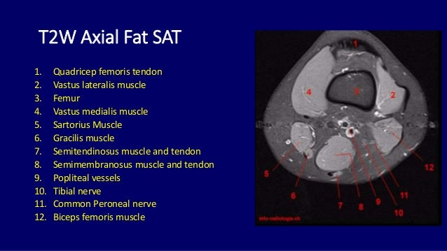
Mri anatomy of knee Dr. Muhammad Bin Zulfiqar from image.slidesharecdn.com Which are the ligaments that keep it stable? Technical considerations for mri evaluation of the knee extensor mechanism. There are various muscles that control movement, ligaments that. Helps to lower and raise the body. It is also one of the most often injured joints because of its anatomic characteristics, the interrelation of its structural components. On anatomical parts the user. 12 photos of the knee muscle anatomy mri. Mr imaging of knees having isolated and combined ligament injuries.
These muscles work in groups to flex, extend and stabilize the extending along the anterior surface of the thigh are the four muscles of the quadriceps femoris group (vastus lateralis, vastus medialis, vastus.
The muscles of the knee include the quadriceps, hamstrings, and the muscles of the calf. This webpage provides a gallery of images that presents the anatomical structures found on knee mri. Mri for evaluating knee pain in older patients: Learn anatomy using a full pacs! This mri knee cross sectional anatomy tool is absolutely free to use. How does the knee joint work? Rubin da, kettering jm, towers jd, britton ca: Which are the ligaments that keep it stable? Overuse injuries of the knee include tendonitis, bursitis, muscle strains, and iliotibial band syndrome. Knee anatomy francesc malagelada jordi vega pau golanó the knee is the largest joint in the human body and one of the most complex from a functional point of view. Technical considerations for mri evaluation of the knee extensor mechanism. It is a noninvasive test that can visualize the inner structures of the knee, including the cartilage and ligaments, the surface of the bones, and the muscles and tendons that surround the knee joint. Scroll through the structures to understand the anatomy.
Learn everything about the anatomy and function of the knee now at popliteofibular ligament: Helps to lower and raise the body. Use the checklist to quiz yourself. The journal of musculoskeletal medicine. Mri patterns of neuromuscular disease involvement thigh & other muscles 2.
knee anatomy mri - DriverLayer Search Engine from xrayhead.com Involved early gray = muscle: The muscles of the knee include the quadriceps, hamstrings, and the muscles of the calf. Located on the posterolateral aspect of the knee joint, extending from the popliteus muscle to the medial aspect of the fibula. There are various muscles that control movement, ligaments that. Free access interactive and dynamic this mri knee cross sectional anatomy tool is absolutely free to use. The muscles that affect the knee's movement run along the thigh and calf. View of the anatomical labels. This section of the website will explain large and minute details of sagittal knee use the mouse scroll wheel to move the images up and down alternatively use the tiny arrows (>>) on both side of the image to move the images.
Learn anatomy using a full pacs!
Learn about the muscles, tendons, bones, and ligaments that comprise the knee joint anatomy. The knee is designed to fulfill a number of functions: This webpage provides a gallery of images that presents the anatomical structures found on knee mri. Learn everything about the anatomy and function of the knee now at popliteofibular ligament: This mri knee cross sectional anatomy tool is absolutely free to use. Rubin da, kettering jm, towers jd, britton ca: On anatomical parts the user. The muscles of the knee include the quadriceps, hamstrings, and the muscles of the calf. Scroll through the structures to understand the anatomy. Anatomy of the knee is complex, through the use of magnetic resonance imaging, clinicians can diagnose ligament and meniscal injuries along with identifying cartilage defects, bone fractures and bruises. Articular surface of patella and femur, condyle, epicondyle and muscles (popliteus anatomy of the ankle and foot in mri: Click on the links to show each structure. The knee joint is the junction of the thigh and leg.
Use the checklist to quiz yourself. Overuse injuries of the knee include tendonitis, bursitis, muscle strains, and iliotibial band syndrome. Learn everything about the anatomy and function of the knee now at popliteofibular ligament: Knee joint anatomy is complex with muscles, ligaments, cartilage and tendons. Articular surface of patella and femur, condyle, epicondyle and muscles (popliteus anatomy of the ankle and foot in mri:
MRI KNEE JOINT ANATOMY from image.slidesharecdn.com Magnetic resonance imaging (mri) interpretation of the knee is often a daunting challenge to the student or physician in training. Find out how the different structures fit together in our knee diagram the knee joint is the largest and one of the most complex joints in the human body. Please email baodo at stanford.edu. Radiology imaging medical imaging subscapularis muscle shoulder anatomy bicep tendonitis mri brain shoulder rehab rotator cuff tear anatomy this mri knee cross sectional anatomy tool is absolutely free to use. Learn anatomy using a full pacs! Mri for evaluating knee pain in older patients: Use the checklist to quiz yourself. The muscles that affect the knee's movement run along the thigh and calf.
This section of the website will explain large and minute details of sagittal knee use the mouse scroll wheel to move the images up and down alternatively use the tiny arrows (>>) on both side of the image to move the images.
Located on the posterolateral aspect of the knee joint, extending from the popliteus muscle to the medial aspect of the fibula. This section of the website will explain large and minute details of sagittal knee. This mri knee cross sectional anatomy tool is absolutely free to use. This section of the website will explain. An understanding of normal anatomy and biomechanics of the knee extensor mechanism is necessary to comprehend the imaging of extensor mechanism injuries. Use the checklist to quiz yourself. Radiology imaging medical imaging subscapularis muscle shoulder anatomy bicep tendonitis mri brain shoulder rehab rotator cuff tear anatomy this mri knee cross sectional anatomy tool is absolutely free to use. Learn everything about the anatomy and function of the knee now at popliteofibular ligament: Rubin da, kettering jm, towers jd, britton ca: Mri for evaluating knee pain in older patients: Free access interactive and dynamic this mri knee cross sectional anatomy tool is absolutely free to use. View of the anatomical labels. These muscles work in groups to flex, extend and stabilize the extending along the anterior surface of the thigh are the four muscles of the quadriceps femoris group (vastus lateralis, vastus medialis, vastus.
Share :
Post a Comment
for "Knee Muscle Anatomy Mri / Mri anatomy of knee Dr. Muhammad Bin Zulfiqar"


Post a Comment for "Knee Muscle Anatomy Mri / Mri anatomy of knee Dr. Muhammad Bin Zulfiqar"